
Stages of Meiosis: Understanding the Division and Phase of Cell Division
Meiosis is a specialized form of cell division essential for sexual reproduction, genetic diversity, and the continuity of life. Unlike mitosis, which produces two genetically identical diploid cells, meiosis reduces the chromosome number by half, resulting in haploid cells that carry a unique combination of genetic material. This process is fundamental for the formation of gametes—sperm and egg cells—and underpins the accurate transmission of genetic information across generations.
The process of meiosis consists of two successive divisions: meiosis I and meiosis II. Each division contains distinct stages and phases that ensure precise chromosome separation and the production of genetically distinct daughter cells. During meiosis I, homologous chromosomes pair and undergo recombination, a process that introduces genetic variation through the exchange of genetic material between maternal and paternal chromosomes. Meiosis II follows without additional DNA replication, separating sister chromatids to produce four haploid daughter cells from a single diploid parent cell. These stages of meiosis demonstrate the intricacy of cellular mechanisms that maintain chromosome integrity while allowing genetic diversity.
Key structural components, including the spindle apparatus, play a critical role in orchestrating chromosome movement during both divisions. Chromosomes align along the equator of the cell during metaphase, are pulled to opposite poles during anaphase, and are enclosed in newly formed nuclei during telophase. Cytokinesis completes each division by physically separating the cytoplasm, resulting in individual haploid cells. Errors during these stages, such as nondisjunction or improper segregation of chromosome pairs, can lead to an abnormal number of chromosomes and are associated with genetic disorders such as Down syndrome, Turner syndrome, and other chromosomal abnormalities.
Meiosis also highlights the contrast between mitosis and meiotic cell division. While mitosis supports growth, tissue repair, and cellular replacement, meiosis is dedicated to sexual reproduction, introducing genetic variation and reducing chromosome number in gametes. The precision of meiosis relies on checkpoints and tightly regulated phases, ensuring that each stage of meiosis progresses correctly and that the resulting cells have the proper set of chromosomes.
By examining the stages of meiosis and their respective phases—prophase, metaphase, anaphase, and telophase—this guide provides a comprehensive view of how chromosomes are duplicated, paired, recombined, and segregated. It also underscores the clinical significance of accurate meiotic division and the consequences of meiotic errors, emphasizing the biological importance of meiosis in human reproduction, heredity, and cellular integrity.
Introduction to Meiosis
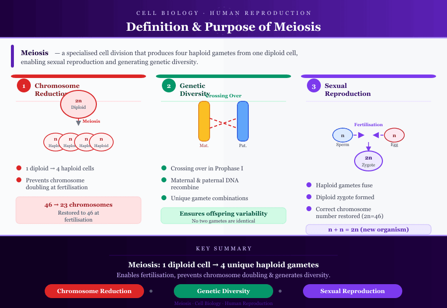
Meiosis is a specialized form of cell division that produces haploid cells necessary for sexual reproduction. Unlike mitosis, which generates two identical diploid cells, meiosis reduces the number of chromosomes in a parent cell by half, resulting in genetically unique haploid daughter cells. This process ensures that gametes—sperm and egg cells—contain a single set of chromosomes, allowing the chromosome number to be restored upon fertilization.
Definition and Purpose of Meiosis in Human Cells
The primary goals of meiosis are:
- Reduction of Chromosome Number:
- One diploid cell divides to produce four haploid cells, each containing half the number of chromosomes as the original.
- This reduction prevents the doubling of chromosomes in the zygote after fertilization.
- Generation of Genetic Diversity:
- Homologous chromosomes undergo pairing during prophase I, allowing crossing over between maternal and paternal chromosomes.
- This recombination produces unique chromosome combinations, ensuring variability among gametes.
- Preparation for Sexual Reproduction:
- Meiosis produces haploid gametes that can fuse during fertilization, forming a diploid zygote with the correct number of chromosomes.
Example:
A human diploid cell has 46 chromosomes (23 pairs). After meiosis, each gamete has 23 haploid chromosomes, half from the mother and half from the father, ready for fertilization.
Importance in Sexual Reproduction
- Ensures Genetic Stability: By halving the chromosome number, meiosis prevents chromosome doubling across generations.
- Promotes Genetic Variation: Independent assortment of homologous chromosomes and crossing over in prophase of meiosis I introduces variation, which is critical for evolutionary adaptation.
- Supports Fertility: Accurate meiotic division is essential to produce viable gametes; errors can lead to infertility or chromosomal abnormalities such as Down syndrome or Turner syndrome.
Overview of Meiosis I and Meiosis II
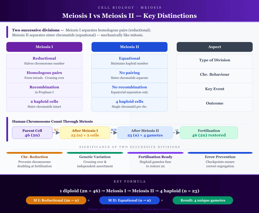
Meiosis is a two-step process of cell division that transforms a single diploid cell into four genetically distinct haploid cells, each containing half the number of chromosomes of the original parent cell. The process occurs in two successive divisions: meiosis I and meiosis II, each with unique roles and characteristics.
Key Distinctions Between Meiosis I and Meiosis II
- Type of Division:
- Meiosis I is the reductional division, reducing the chromosome number by half. It separates homologous chromosome pairs so that each daughter cell receives only one chromosome from each pair.
- Meiosis II is the equational division, where sister chromatids are separated without changing the haploid chromosome number. Mechanistically, meiosis II is similar to mitosis.
- Chromosome Behavior:
- In meiosis I, homologous chromosomes form tetrads and may exchange segments via recombination/crossing over during prophase I.
- In meiosis II, no pairing occurs. Each haploid cell contains unpaired chromosomes, and the division separates sister chromatids along the equator of the cell.
- Outcome:
- Meiosis I produces two haploid cells, each with duplicated chromosomes (sister chromatids intact).
- Meiosis II generates four haploid daughter cells, each with a single chromatid per chromosome, ready for fertilization.
Chromosome Number in Parent Cells vs Daughter Cells
- Parent Cell:
- Begins as a diploid cell (2n) with two sets of chromosomes, one maternal and one paternal.
- Each chromosome consists of sister chromatids, connected at a centromere.
- After Meiosis I:
- Two haploid cells are produced, each containing one chromosome from each homologous pair.
- Chromosomes still have sister chromatids, maintaining a temporary duplication.
- After Meiosis II:
- Four haploid daughter cells are produced, each containing a single set of chromosomes.
- This ensures gametes have half the number of chromosomes as the original parent cell, maintaining genetic stability.
Example:
A human diploid cell has 46 chromosomes. After meiosis I, each cell has 23 chromosomes with sister chromatids. After meiosis II, four haploid gametes are produced, each with 23 single chromatids, ready for sexual reproduction.
Significance of Two Successive Divisions
- Reduction of Chromosome Number:
- Ensures gametes contain half the number of chromosomes, preventing doubling at fertilization.
- Generation of Genetic Variation:
- Crossing over in prophase I and independent assortment during metaphase I create diverse chromosome combinations, essential for evolution.
- Preparation for Fertilization:
- Produces haploid gametes capable of fusing to restore diploid chromosome number.
- Error Prevention:
- Sequential divisions allow checkpoints to ensure correct alignment, recombination, and segregation of chromosomes.
Stages of Meiosis I
Meiosis I, also called the first meiotic division, is the reductional division, reducing the diploid chromosome number to haploid. It sets the stage for genetic variation and ensures that each daughter cell receives one chromosome from each homologous pair.
Prophase I
Prophase I is the longest and most complex phase of meiosis, characterized by several critical events:
- Homologous Chromosome Pairing (Synapsis):
- Each diploid parent cell contains a pair of homologous chromosomes (one maternal and one paternal).
- These chromosomes align closely along their lengths to form tetrads (groups of four chromatids).
- Synapsis is facilitated by the synaptonemal complex, a protein structure that stabilizes paired chromosomes.
- Recombination/Crossing Over and Genetic Variation:
- During synapsis, non-sister chromatids may exchange segments of DNA in a process called crossing over.
- This recombination produces new combinations of maternal and paternal genes, contributing to genetic variation in the resulting haploid cells.
- Example: If a gene for eye color is on a maternal chromosome and a gene for hair color is on the paternal chromosome, crossing over can create a gamete carrying the maternal eye color gene and paternal hair color gene together.
- Comparison to Mitotic Prophase:
- In mitosis, chromosomes condense but do not pair, and recombination does not occur.
- Meiosis I prophase is unique for homologous pairing and genetic recombination, which are absent in mitosis.
Metaphase I
- Alignment of Homologous Chromosomes at the Metaphase Plate:
- Tetrads align along the equator of the cell.
- Each homologous chromosome is oriented randomly, contributing to independent assortment and genetic diversity.
- Role of Spindle Fibers in Chromosome Movement:
- Spindle fibers attach to centromeres via kinetochores.
- They help orient the chromosomes so that each daughter cell will receive one chromosome from each homologous pair.
- Correct attachment is crucial to prevent errors in meiosis, such as nondisjunction, which can cause disorders like Down syndrome.
Anaphase I
- Separation of Homologous Chromosomes:
- The spindle fibers pull homologous chromosomes toward opposite poles of the cell.
- Unlike mitosis, sister chromatids remain attached at their centromeres during this division.
- Effect on Chromosome Number:
- The cell transitions from diploid (2n) to haploid (n) because each pole receives only one chromosome from each homologous pair, though each still contains two chromatids.
Telophase I and Cytokinesis
- Nuclear Envelope Reformation:
- Chromosomes may decondense slightly, and a nuclear envelope may form around each set of chromosomes.
- Formation of Two Haploid Daughter Cells:
- Cytokinesis divides the cytoplasm, resulting in two haploid cells.
- Each cell has a unique set of chromosomes due to crossing over and independent assortment.
Stages of Meiosis II
Meiosis II, the second meiotic division, is the equational division. It resembles mitosis, as it separates sister chromatids in haploid cells, resulting in four genetically distinct haploid daughter cells.
Prophase II
- Preparation for the Second Division:
- Chromosomes condense again, and the spindle apparatus reforms.
- Each chromosome still consists of two sister chromatids.
- Comparison to Prophase I and Mitosis:
- No pairing or crossing over occurs in prophase II, unlike prophase I.
- The process is similar to mitotic prophase, except the cells are haploid.
Metaphase II
- Chromosomes Align Along the Metaphase Plate:
- Individual chromosomes (not homologous pairs) line up along the equator of the cell.
- Differences from Metaphase I:
- In metaphase I, tetrads align; in metaphase II, sister chromatids align individually.
- This ensures that each haploid daughter cell receives one chromatid from each chromosome.
Anaphase II
- Separation of Sister Chromatids:
- Centromeres split, and spindle fibers pull sister chromatids to opposite poles of the cell.
- Chromosome Movement Toward Poles:
- Each chromatid becomes an independent chromosome, ensuring proper haploid composition in the resulting cells.
Telophase II and Cytokinesis
- Completion of Nuclear Division:
- Nuclear envelopes form around each set of chromosomes.
- Production of Four Haploid Daughter Cells:
- Cytokinesis divides the cytoplasm.
- The result is four genetically distinct haploid cells, each with one complete set of chromosomes.
- These cells are ready to participate in fertilization, restoring the diploid chromosome number in the zygote.
Clinical Relevance for Sexual Reproduction
- Accurate meiotic division ensures that gametes have the correct chromosome number.
- Genetic variation from crossing over and independent assortment is fundamental for species diversity and adaptation.
- Errors in meiosis, such as nondisjunction, can produce aneuploid gametes, leading to genetic disorders:
- Trisomy 21 (Down syndrome) – extra chromosome 21
- Turner syndrome – missing X chromosome in females
- Klinefelter syndrome – extra X chromosome in males
- Understanding the stages of meiosis is essential in genetics, reproductive biology, and clinical practice, especially in fertility studies and genetic counseling.
Meiosis vs Mitosis: Key Differences
Understanding the differences between meiosis and mitosis is fundamental for comprehending cell division processes, chromosome dynamics, and clinical applications in nursing. While both processes involve nuclear division, they serve distinct biological purposes and produce different outcomes.
Differences in Stages, Chromosome Number, and Type of Cell Division
- Purpose of Division:
- Mitosis is a type of cell division aimed at producing genetically identical cells for growth, tissue repair, and cell replacement.
- Meiosis is a meiotic cell division designed to generate haploid cells for sexual reproduction, ensuring that offspring inherit a unique combination of maternal and paternal chromosomes.
- Number of Divisions:
- Mitosis involves one division resulting in two daughter cells.
- Meiosis includes two successive divisions—meiosis I and meiosis II—producing four haploid daughter cells from a single diploid parent cell.
- Chromosome Number in Daughter Cells:
- Mitosis: Daughter cells are diploid, containing the same number of chromosomes as the parent cell (2n → 2n).
- Meiosis: Daughter cells are haploid, containing half the number of chromosomes compared to the parent cell (2n → n).
- Stages and Key Events: Feature Mitosis Meiosis Prophase Chromosomes condense; no homologous pairing; spindle forms Prophase I: homologous chromosomes pair (synapsis), crossing over occurs; Prophase II: spindle reforms in haploid cells Metaphase Chromosomes align along metaphase plate Metaphase I: homologous chromosome pairs align; Metaphase II: sister chromatids align Anaphase Sister chromatids separate to opposite poles Anaphase I: homologous chromosomes separate; Anaphase II: sister chromatids separate Telophase & Cytokinesis Nuclear envelope reforms; two diploid daughter cells produced Telophase I: two haploid cells; Telophase II: four haploid daughter cells produced Genetic Variation Daughter cells genetically identical Daughter cells genetically distinct due to crossing over and independent assortment
- Type of Chromosome Separation:
- Mitosis: Separates sister chromatids, producing identical copies.
- Meiosis I: Separates homologous chromosomes, reducing chromosome number.
- Meiosis II: Separates sister chromatids, similar to mitosis but in haploid cells.
- Occurrence of Genetic Recombination:
- Mitosis: No recombination occurs; cells are clones.
- Meiosis: Crossing over in prophase I and independent assortment in metaphase I create genetic variation, crucial for sexual reproduction.
Example:
A human somatic cell undergoing mitosis maintains 46 chromosomes in both daughter cells. In contrast, a germ cell undergoing meiosis produces gametes with 23 chromosomes each, ensuring that fertilization restores the diploid number. Crossing over ensures that each gamete is genetically unique.
Clinical Relevance for Nursing Practice and Exams
- Understanding Genetic Disorders:
- Mistakes during meiosis such as nondisjunction can result in disorders like:
- Down syndrome (trisomy 21)
- Turner syndrome (monosomy X)
- Klinefelter syndrome (XXY)
- Nurses must understand these processes to provide patient education and support genetic counseling.
- Mistakes during meiosis such as nondisjunction can result in disorders like:
- Application in Reproductive Health:
- Knowledge of meiosis is essential for understanding fertility, gamete formation, and assisted reproductive technologies.
- Distinguishing Cell Types in Pathology:
- Differentiating somatic cells undergoing mitosis versus germ cells undergoing meiosis helps in understanding tumor formation, tissue regeneration, and cell differentiation.
- Exam Relevance:
- Nursing students are often tested on chromosome behavior, haploid vs diploid cells, and phase-specific events, particularly in sexual reproduction and genetics modules.
- Visualizing key differences in stages, chromosome number, and type of cell division aids in answering exam questions on meiosis vs mitosis accurately.
Example:
In a patient with suspected genetic abnormalities, understanding that errors during meiosis can lead to aneuploidy allows nurses to anticipate clinical signs, prepare for appropriate counseling, and collaborate with genetic specialists.
Errors in Meiosis and Clinical Implications
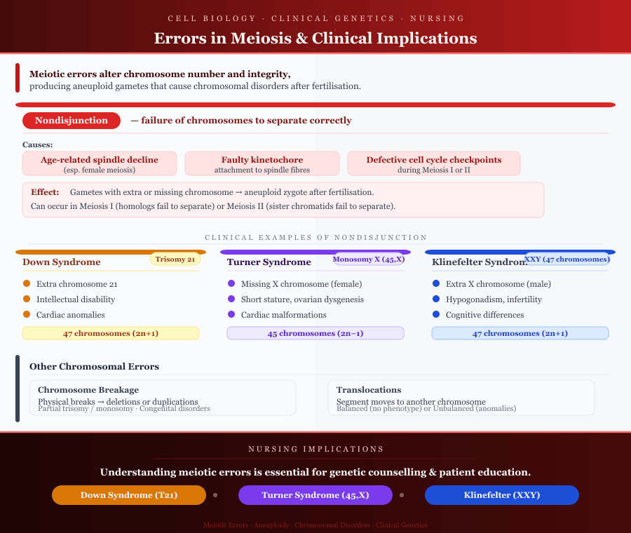
Errors during meiotic division can have profound consequences on chromosome number, genetic integrity, and ultimately, patient outcomes. Understanding these errors is essential for clinical nursing practice, genetic counseling, and patient education.
Common Meiotic Errors
1. Nondisjunction
- Definition: Nondisjunction occurs when homologous chromosomes in meiosis I or sister chromatids in meiosis II fail to separate properly, resulting in gametes with an abnormal number of chromosomes.
- Causes:
- Age-related decline in meiotic spindle function, particularly in female meiosis.
- Faulty kinetochore attachment to spindle fibers.
- Defective cell cycle checkpoints during the first or second meiotic division.
- Clinical Examples:
- Down syndrome (Trisomy 21): An extra chromosome 21 due to nondisjunction. Symptoms include intellectual disability, characteristic facial features, and cardiac anomalies.
- Turner syndrome (Monosomy X): Missing X chromosome in females; leads to short stature, ovarian dysgenesis, and cardiac malformations.
- Klinefelter syndrome (XXY): Extra X chromosome in males; results in hypogonadism, infertility, and cognitive differences.
- Effect on Cell Division:
- Produces gametes with one extra or one missing chromosome, which, after fertilization, leads to an aneuploid zygote.
- Highlights the importance of accurate spindle assembly and chromosome segregation during meiosis.
2. Other Chromosomal Errors
- Chromosome Breakage:
- Physical breaks in chromosomes during prophase of meiosis can lead to deletions or duplications.
- Can result in partial trisomy or monosomy, contributing to congenital disorders.
- Translocations:
- A segment of a chromosome breaks and attaches to another chromosome.
- May be balanced, with no immediate phenotype, or unbalanced, causing developmental anomalies or infertility.
- Example:
- Robertsonian translocation can lead to familial Down syndrome, where chromosome 21 is translocated to another chromosome, producing gametes with abnormal chromosome pairs.
Cellular Checkpoints and Error Prevention
Mechanisms to Reduce Mistakes in Meiosis:
- Spindle Assembly Checkpoint (SAC):
- Ensures all chromosomes align at the metaphase plate before anaphase begins.
- Detects improperly attached spindle fibers to kinetochores, preventing premature separation.
- DNA Damage Checkpoints:
- Monitor chromosome integrity during prophase I, particularly during crossing over.
- Recombination errors or double-strand breaks activate repair mechanisms before progression to metaphase I.
- Apoptotic Pathways:
- Cells with severe meiotic errors may undergo programmed cell death, preventing abnormal gametes from contributing to fertilization.
- Cohesin Proteins:
- Maintain sister chromatid cohesion until the proper phase of division, reducing the risk of premature separation.
Example:
- If a chromosome fails to attach to the spindle during metaphase I, the spindle assembly checkpoint halts progression until the error is corrected, ensuring accurate chromosome number in daughter cells.
Nursing Implications of Meiotic Errors
- Understanding Abnormal Chromosome Numbers in Patients:
- Knowledge of meiotic errors helps nurses anticipate clinical presentations of aneuploidy.
- Recognizing signs of disorders like Down syndrome or Turner syndrome informs patient care plans and monitoring requirements.
- Genetic Counseling and Patient Education Relevance:
- Nurses play a key role in educating patients and families about the genetic basis of disorders arising from meiosis errors.
- Counseling includes:
- Explaining the process of meiotic division and how errors occur.
- Discussing the probability of recurrent chromosomal abnormalities.
- Informing patients about screening, prenatal testing, and fertility options.
- Clinical Application in Nursing Practice:
- Incorporating knowledge of meiotic errors aids in:
- Planning prenatal care for at-risk pregnancies.
- Supporting families with genetic conditions.
- Collaborating with geneticists and reproductive specialists.
- Incorporating knowledge of meiotic errors aids in:
Example:
- A nurse explaining trisomy 21 to expectant parents can describe how a nondisjunction event in maternal meiosis results in an extra chromosome 21, emphasizing why early detection via screening is important.
Conclusion
Understanding the stages of meiosis is fundamental for comprehending how cell division drives genetic variation and supports sexual reproduction. Through the two successive divisions—meiosis I and meiosis II—a single diploid parent cell gives rise to haploid daughter cells, each containing a unique set of chromosomes. The precise orchestration of prophase, metaphase, anaphase, telophase, and cytokinesis ensures accurate chromosome segregation and maintains genomic stability.
Key distinctions between meiosis and mitosis highlight the specialized role of meiotic division in producing genetically diverse gametes, contrasting with the uniformity of mitotic cell division used for growth and repair. The processes of homologous chromosome pairing, recombination, and independent assortment are essential mechanisms that generate variation, which underpins evolution and adaptability.
Errors during meiosis, such as nondisjunction, chromosome breakage, and translocations, can disrupt the number and structure of chromosomes, leading to clinical conditions like Down syndrome, Turner syndrome, and Klinefelter syndrome. Cellular safeguards, including spindle assembly checkpoints, DNA repair mechanisms, and apoptotic pathways, work to reduce the occurrence of these errors, emphasizing the complexity and precision of meiotic processes.
From a clinical perspective, a strong understanding of meiotic stages and errors equips healthcare professionals with the knowledge to:
- Interpret and explain genetic conditions to patients and families.
- Support genetic counseling and patient education initiatives.
- Recognize the importance of chromosome integrity in reproductive health and fertility assessments.
In summary, meiosis is more than just a sequence of cellular events—it is a foundational process that ensures the continuity, diversity, and integrity of life. Mastery of its stages, mechanisms, and potential errors not only strengthens scientific understanding but also enhances the ability to translate this knowledge into practical, patient-centered care.
Frequently Asked Questions
What are the stages of meiosis in order?
Meiosis consists of two successive divisions, each with distinct phases:
- Meiosis I (First Meiotic Division – Reductional Division):
- Prophase I – Homologous chromosome pairing (synapsis) and crossing over
- Metaphase I – Tetrads align at the metaphase plate
- Anaphase I – Homologous chromosomes separate to opposite poles
- Telophase I – Nuclear envelopes reform
- Cytokinesis – Two haploid daughter cells are produced
- Meiosis II (Second Meiotic Division – Equational Division):
- Prophase II – Chromosomes condense; spindle forms
- Metaphase II – Chromosomes align individually at the equator of the cell
- Anaphase II – Sister chromatids separate to opposite poles
- Telophase II – Nuclear envelopes reform
- Cytokinesis – Four genetically distinct haploid cells are produced
Why is Prophase I complex?
Prophase I is the longest and most intricate phase of meiosis because:
- Homologous chromosomes pair (synapsis) to form tetrads.
- Crossing over/recombination occurs, exchanging genetic material between maternal and paternal chromosomes, creating genetic variation.
- The synaptonemal complex facilitates chromosome alignment and ensures proper segregation.
- Unlike mitotic prophase, these events are unique to meiosis and essential for producing haploid gametes.
Difference between Meiosis I and II
| Feature | Meiosis I | Meiosis II |
|---|---|---|
| Type of division | Reductional – reduces chromosome number | Equational – separates sister chromatids |
| Chromosomes separated | Homologous chromosomes | Sister chromatids |
| Resulting cells | Two haploid daughter cells, each with duplicated chromatids | Four haploid daughter cells, each with single chromatids |
| Genetic variation | Crossing over occurs in Prophase I | No crossing over occurs |
| Similarity to mitosis | Different from mitosis due to homologous pairing | Similar to mitosis, but in haploid cells |
When does genetic variation occur?
Genetic variation occurs primarily during Prophase I of meiosis through:
- Crossing over (recombination) – exchange of DNA between non-sister chromatids of homologous chromosomes.
- Independent assortment in Metaphase I – random orientation of homologous chromosome pairs at the equator of the cell results in different combinations in haploid daughter cells.
These processes ensure that gametes are genetically unique, contributing to diversity in sexual reproduction.